
6 auxiliary traction technology: let endoscopic mucosal stripping become simple
Endoscopic mucosal resection (ESD) has become a widely accepted minimally invasive surgical procedure for the treatment of early carcinoma of the gastrointestinal tract due to its high complete resection rate and good safety. The basic procedure is divided into three steps: the submucosal injection, the annular incision of the lesion and the submucosal tissue of the lesion.
ESD treatment of difficult parts of the lesion, usually take repeated submucosal injection, although the subsequent stripping operation provides a limited view of the operation, but the operation time is long, increase the risk of bleeding, perforation. Another simple solution is to adjust the patient's body position according to the direction of gravity, so that the stripping surface to obtain the desired tension, and provide a good operation field of vision, but the patient can choose a limited body position, limit the use of results.
Therefore, how to achieve a good "vision" in the operation, to achieve a safe, accurate cutting and stripping operation method is the key to solve the above problems.
Inspired by the surgical technique, all kinds of endoscope assisted traction technology came into being. According to can be roughly divided into traction for the in vivo and in vitro traction traction force source location. According to the traction device can be divided into metal clip silk combined traction technology, metal clip circle of elastic joint traction technology, S-O metal clip traction technology, magnetic anchor technology etc..
This paper summarizes the advantages and disadvantages of various relevant traction technology, in order to provide reference for clinical choice.
Combined traction technology of metal wire clip
At present, one of the most widely used methods in the esophagus, stomach, duodenum and colon surgery are applied. The technical points are:
1 in vitro ligation of the silk thread in the metal clip between the two clamping arms, through the endoscopic treatment channel, the metal clip is fixed on the cut edge of the lesion (Figure 1a). Light pull the thread to maintain appropriate tension, so that the lesion was fully carried, and then the implementation of a complete stripping. But because of its limited traction direction, it is difficult to apply force to the side of the lesion.
2. Later, scholars have improved on the method, namely in the first metal clip on the side wall of the mucous membrane fixed on another metal clips, the silk thread ligation in the first metal clip from bypass, produced similar to the "block" effect, in order to obtain the anal side direction tension (Fig. 1B).
3 another more direct way is that the second metal clips and the thread is fixed on the side of the mucosa, so that the peel tissue from the side of the tension, see Figure 1c. But this method is insufficient, one is silk itself can only provide directional pull; second is silk itself does not shrink, with the increase of the release face, the thread tension decreases, its role will be decreased or disappeared.
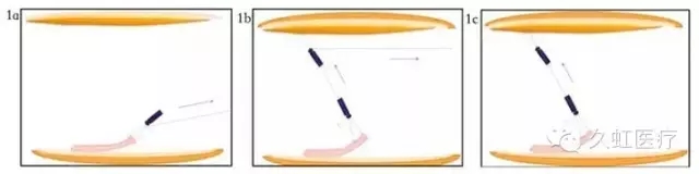
Figure 1 different variants of the metal wire clip combined traction technology (1a. direct wire traction method; 1b. using the "pulley group" effect of the wire traction method; 1c. on the side of the wire traction method)
The principle of the method is simple, operation better, material is relatively easy to obtain, thread, nylon thread, floss can be as wire material. A randomized controlled study showed that, compared with the traditional ESD, it shortened the operation time, and reduced the number of submucosal injection, but in the operation process may have the problem of metal clip off.
Combined traction technology of metal elastic ring
Metal clip circle of elastic joint traction technology to a body traction technology, relative to the cable, the elastic ring on its extension can be in vivo provides a force. Because the external force is not needed, the restriction of the anatomical position and the lumen size of the alimentary canal is not suitable for the difficult position of the ESD operation.
The material used by the elastic ring has been widely used in clinical, such as O type loop of esophageal vein curve, surgical sterile gloves, etc., in the in vivo nature is stable, will not produce allergic reaction.
Actual operation, the in vitro to 3.0 thread will medical elastic ring ligation in the metal clip on the side of the arm, then with metal clip incorporated together after the release sheath (Figure 2a); in vivo completed pre separated mucosa, the metal clip is fixed on the edge of the lesion and the second metal clip side arm through an elastic ring is fixed on the lesion on the edge of the side (Figure 2b-c); surface mucosa lesions gradually because of the action of the elastic force of the valgus, the exposure field of view (Figure 2D).
After the operation, the auxiliary instrument was recovered. Considering the elastic force ring to advance to the intrathecal and late and easy operation, this circle, the folding and unfolding of the ideal radius is divided into 2 mm and 5 mm, silk thread ligation is red, to operation background contrast.
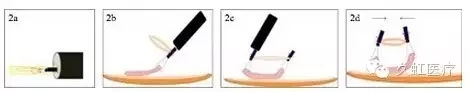
Figure 2 metal clip wire combined with traction technology flow chart (2a. Metal clip and an elastic ring in vitro storage to release the sheath; figure 2B. Accompanied by elastic ring first gold metal clip in the pre separation mucosa; figure 2C. A second gold metal clip through the elastic ring is fixed in the contralateral separated mucosa; figure 2D. Two pieces of metal clip on elastic force turned up mucosal exposure field)
S-O metal clamp traction technology
In 2009, two Japanese scholars based on the spring, metal clips and nylon ring for the design of another in vivo traction technology, and with the first letter of the two names of the first letter S-O metal clip traction technology. The method consists of a metal clip elastic ring combined with traction of the evolvement, to spring instead of the elastic force ring, trying to obtain greater scalability, to adapt to large, ESD shallow early colon tumor resection (Figure 3).
The spring length of the device is 7 mm, the width is 1.8 mm, the force of 1 G above can make the spring deformation, the maximum extension length is 10 times, the maximum can be 20 G. After the completion of the mucosal stripping, with endoscopic scissors to open the nylon loop, with the specimen removed. Clinical prospective controlled study showed that the operation time of the experimental group, the tumor resection rate and the safety of the experimental group were better than those of the control group.
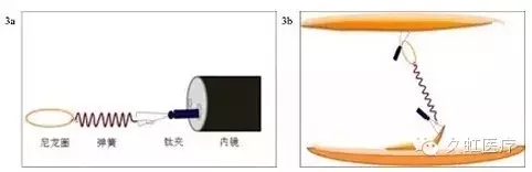
Figure 3 S-O metal clip composition and drawing mode (S-O 3a. metal clip structure diagram; S-O 3b. metal clip pull pull mode)
Magnetic force anchor traction technology
Depending on the magnetic field generated by the magnetic force for ESD operation provides a different angle, direction and size of the traction force is a very creative idea, a small magnet bundled ligation line in a metal clip or two metal sandwiched between, to single / dual channel endoscope in the fixed position, in vitro another piece of magnet with the magnetic force is generated, of the stripping process implementation of stretch.
This technique has been improved since its invention. Magnet type can be divided into magnet, SmCo permanent magnet and neodymium rare earth magnet, while the latter is the strongest magnetic force of magnetic material, is in the magnet surface can prevent human tissue to nickel allergy. In vitro magnet shape can be square, cylindrical or saucer shaped, while in vivo magnets are easy to tie up the ring. The magnet anchor can be divided into single and double anchor anchor, the latter more open view (Figure 4).
Need to pay attention to, because between the magnet magnetic size usually with the volume and distance dependent, row posterior wall of gastric body ESD procedure should increase the volume or number of in vitro and in vivo magnet, but amount of magnets increase may impede the operation field.
At present, magnetic anchor traction technology applied in vitro magnet have been equipped with standard can be telescopic arm (Japan SFC Co Ltd, FA-M-VC2) to reduce the workload of the assistant, the platform assembly time average is only 4 minutes, the operation time is shorter than that of the control group. But the price of the whole system is more expensive, and the related research is limited to the stomach of the animal, which needs further study.
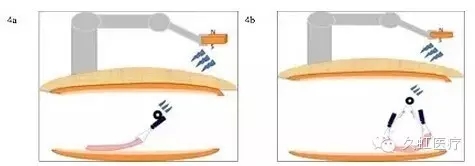
Figure 4 magnetic anchor traction technology of different variants (4a. The use of single anchor magnetic anchor traction technology flow chart; 4B using double anchor magnetic anchor traction technology flow chart, due to the formation of plane triangle, the magnet is at its top, so far away from the plane of dissection, can provide a more broad perspective)
Clamp traction technology
As an in vitro traction technology, the traction technique has its own advantages:
1 with the characteristics of hard materials, you can target the organization to implement the "pull", "push", "pick" and "rotation" and other techniques can not match the many kinds of action, to achieve the best results;
2 can be replaced and repeatedly pull point clamp organization, operation and assistant operation intention reached;
3 withdrawal is convenient, and can help the recovery of surgical specimens.
Traction forceps under the guidance of the endoscope into a fixed position, using single channel endoscope can be attached to the lens body, with self-made sheath or another built-in in the biopsy channel of the pincers fixed (Figure 5a-b) in the clinical operation, to achieve the first step and first step. Such as the use of double channel endoscope, through which a biopsy channel into the implementation of pull, the other channel into the electric knife to implement the stripping operation (Figure 5C).
 TOP
TOP